Rahul Sarma,
Minakshi Das,
Tiluttama Mudoi,
Kaustav Kalyan Sharma,
Jibon Kotoky,
Rajlakshmi Devi ![]()
For correspondence:- Rajlakshmi Devi Email: rajiasst@gmail.com Tel:+3612279939
Received: 1 June 2015 Accepted: 11 December 2015 Published: 29 January 2016
Citation: Sarma R, Das M, Mudoi T, Sharma KK, Kotoky J, Devi R. Evaluation of antioxidant and antifungal activities of polyphenol-rich extracts of dried pulp of Garcinia pedunculata Roxb and Garcinia morella Gaertn (Clusiaceae). Trop J Pharm Res 2016; 15(1):133-140 doi: 10.4314/tjpr.v15i1.19
© 2016 The authors.
This is an Open Access article that uses a funding model which does not charge readers or their institutions for access and distributed under the terms of the Creative Commons Attribution License (http://creativecommons.org/licenses/by/4.0) and the Budapest Open Access Initiative (http://www.budapestopenaccessinitiative.org/read), which permit unrestricted use, distribution, and reproduction in any medium, provided the original work is properly credited..
Purpose:To evaluate the antioxidant and antifungal activities of polyphenol-rich extracts of the dried fruit pulp of Garcinia pedunculata (GP) and Garcinia morella (GM) to determine their traditional claims of therapeutic activity against certain diseases.
Methods:Analysis of total phenolic (TP) and flavonoid (TF) contents of the extracts were performed by Folin-Ciocalteau and Arvouet-Grand methods. The antioxidant activity of the extracts was determined by 1,1-diphenyl-2-picrylhydrazyl (DPPH), hydrogen peroxide (H2O2) free radical scavenging activity, reducing power and in vitro lipid peroxidation (LPO). Antifungal activity was evaluated by agar-well diffusion method while mineral content was evaluated by atomic absorption spectrophotometry (AAS).
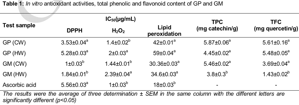
Results:Significant amounts of TP (5.87 ± 0.06 and 5.46 ± 0.02 mg catechin eqivalents/g) and TF (5.61 ± 0.16 and 3.69 ± 0.04 mg quercetin equivalents/g) were found in the cold water (CW) extracts of GP and GM, respectively, along with DPPH free radical scavenging activity (50 % inhibitory concentration (IC50) = 3.53 ± 0.04 and 1 ± 0.03μg/mL) and H2O2-radical scavenging activity (IC50 = 1.4 ± 0.02 and 1.44 ± 0.01 μg/mL). Results indicated that the CW extracts of GP and GM were potent reducing agent than the HW extracts. CW extract of both species prevented in vitro LPO (IC50= 42 ± 0.01 and 30.36 ± 0.03 μg/mL) significantly. The antifungal activity of GP and GM extracts against some human dermatophytes was high. High concentrations of K and Fe were found in the extracts.
Conclusion:GP and GM extracts have great potential as a source for useful antioxidant and antifungal agents.
Introduction
Reactive oxygen species (ROS) such as superoxide anion (O-2), perhydroxy radical (HOO-), hydroxyl radical (HO-) and nitric oxide (NO) radical are generated during the process of cellular oxidation. These electrically charged radicals are highly unstable, reactive in nature and easily attack proteins, nucleic acid, mitochondria and enzymes in biological systems resulting damage in the cell. High levels of reactive free radicals and oxygen species create oxidative stress, which leads to detrimental effects, including lipid peroxidation (LPO) of cellular membranes, alteration of lipid–protein interaction, enzyme inactivation, DNA breakage, and eventually the promotion of mutations that initiate tumor progression [1,2].
Oxidative damage caused by free radicals and reactive oxygen species contribute to more than one hundred disorders in humans including atherosclerosis, arthritis, ischemia and reperfusion injury of many tissues, central nervous system injury, gastritis, diabetes and cancer [3,6]. Antioxidant plays an important role in protecting health from free radicals and reactive oxygen species. It is therefore important to evaluate the consumption of those fruits and vegetables which have been traditionally claimed for having therapeutic values.
GP and GM (family: Clusiaceae) are two popular indigenous fruits of NE region of India locally known as Bor thekera and Kuji thekera respectively. The mature fruit is eaten cooked or raw; generally it is mixed with other vegetables, especially with pulses. Dried pulp of these fruits is used as an antiscorbutic, astringent, cooling, cardiotonic, emollient, antidiarrhoeic, antidysentric, indyspepsia and in flatulence [7]. A field survey was carried out in different parts of Assam to collect the views of different people about the use of GP and GM fruits whereby maximum people claimed the use of dried pulp of the fruit is having therapeutic value than the consumption of raw fruits [8]. In this regard, the dried pulps of the fruit tested on cholesterol fed mice in our laboratory and it showed the hypolipidemic and anti-atherosclorotic effect [9].
There are hardly any reports available on antioxidant and antifungal activity of the GP and GM fruits of NE region of India. So, this study was designed to determine the antioxidant and antifungal activity of polyphenolic enriched extracts of GP and GM fruits.
Methods
Plant material
The fruits GP and GM were collected from local markets of Assam, India in the month of April 2013 with authentication by the Botanist of Gauhati University, Guwahati, Assam and a voucher specimens (IASST/LSD/PM-11 and IASST/LSD/PM-12) were deposited at the medicinal and aromatic plant section, Life Sciences Department, Institute of Advanced Study in Science and Technology (IASST), Assam, India. The fruits were cut into small pieces, sundried for 10-15 days and kept at room temperature for further analysis.
Preparation of cold and hot water extracts
Cold water (CW) extracts of dried pulp of GP and GM were prepared by soaking 5 g of dried pulp in 100 mL drinking water. The mixture was stirred vigorously and allowed to stand for 24 h at room temperature. Thereafter, it was filtered by Whatman filter paper and stored at 4 oC for further use. For hot water (HW) extraction, 5 g of dried pulp each of GP and GM were kept in 100 mL of drinking water and the samples were boiled for 30 min and added into a conical flask and agitated on a rotary shaker for 48 h. Then it was filtered by Whatman filter paper (diameter 125 mm) and filtrate was stored at 4 oC for analysis of different parameters.
Determination of total phenolic content (TPC)
TPC of dried pulp extract of GP and GM was determined by Folin-Ciocalteu method with slight modification [10] Briefly, 0.5 mL of extract was mixed with Folin-Ciocalteu reagent (2.5 mL, diluted 10 times) and incubated for 2 min at room temperature followed by addition of sodium carbonate solution (2 mL, 7.5 % w/v). The mixture was then allowed to stand for 30 min at room temperature and absorbance was measured at 765 nm. TPC was calculated as a catechin equivalence from the calibration curve of catechin standard solutions and expressed as mg catechin/g dried pulp of the sample.
Evaluation of total flavonoid content (TFC)
TFC was estimated according to the Dowd method as adapted by Arvouet-Grand et al [11]. Briefly, 2 mL of extract was mixed with 2 mL of aluminiumtrichloride (AlCl3) in methanol (2 %). The absorbance was read at 415 nm after 10 min. Quercetin was used as reference compound and the results were expressed as mg of quercetin equivalence (QE)/g of dried pulp of fruit.
Evaluation of 2, 2-diphenyl-1-picrylhydrazyl (DPPH) radical scavenging activity
The free radical scavenging ability of the extracts against DPPH free radical was evaluated as described by DPPH method [12]. The solution of DPPH in methanol (6 × 10⁻⁵ M) was prepared just before UV measurements. Samples were added to DPPH solution in 1:1 ratio followed by vortexing. The reaction was allowed to take place in the dark at room temperature. The absorbance at 515 nm was measured at different time intervals. Ascorbic acid served as a standard. IC50 value (the concentration required to scavenge 50 % of the free radical) was estimated from a plot of % inhibition against concentrations of the sample solutions. Scavenging activity (S) was calculated as Eq 1.
S (%) = {(Ac – As)/Ac} 100…………………. (1)
where Ac and As are the absorbance of control and sample, respectively.
Determination of hydrogen peroxide (H2O2) scavenging activity
The scavenging assay for H2O2 was performed by a standard method [13]. A 43 mM solution of H2O2 was prepared in 0.1 M phosphate buffer solution (pH 7.4). Samples (1 mL) were mixed with 43 mM H2O2 solution (0.6 mL). After 10 min, the reaction mixture absorbance was determined at 230 nm. The phosphate buffer without hydrogen peroxide was used as a blank. Ascorbic acid was used as a reference compound. H2O2-scavenging activity (H) of calculated as in Eq 2.
H (%) = {(Ac – As)/Ac} 100 ………………. (2)
where Ac and As are the absorbance of control and sample, respectively.
Assessment of reducing power activity
Reducing power of samples was determined by ferric reducing power assay [14]. Briefly,2.5 mL of 0.2 M phosphate buffer (pH 6.6) and 2.5 mL of 1 % potassium ferricyanide were added to 1 mL sample solution and mixed gently. The mixtures were incubated at 50 oC in a water bath for 20 min. Reaction was stopped by adding 2.5 mL of 10 % trichloroacetic acid (TCA) and the mixtures were centrifuged at 4000 rpm for 10 min. From the top layer, 2.5 mL was transferred into tubes containing 2.5 mL distilled water and 0.5 mL of 0.1 % ferric chloride (FeCl3.6H2O). The resulting solutions were mixed well and after 5 min the absorbance was measured at 700 nm against blanks.
In vitro lipid peroxidation (LPO)
LPO induced by Fe2+-ascorbate system in rat liver homogenate [15], was estimated as thiobarbituric acid reacting substances (TBARS) [16]. The reaction mixture contained rat liver homogenate 0.25 mL (10 % w/v in 0.05 M phosphate buffer, pH 7.4), 0.1 mL tris-HCl buffer (150 mM, pH 7.2), 0.05 mL ascorbic acid (0.1 mM), 0.05 mL FeSO4.7H2O (4 mM) and 0.05 mL of fruit extract. The mixture was incubated at 37 oC for 1 h and then 1.5 mL 2-thiobarbituric acid (TBA, 0.8 % w/v), 1.5 mL acetic acid (20 %) and 0.2 mL sodiumdodecyl sulfate (SDS, 8.1 % w/v) were added to the reaction mixture. The mixture was made up to 4.0 mL with distilled water and heated at 95 oC for 60 min. After cooling with tap water, 1.0 mL distilled water and 5.0 mL of a mixture of n-butanol and pyridine (15:1, v/v) were added. The mixture was shaken vigorously and centrifuged at 5000 rpm for 10 min. After centrifugation, the optical density of the butanol layer was measured at 532 nm.
Assay for antifungal activity
The screening of antifungal activity of the extracts was determined by employing agar well diffusion method [17,18].
Preparation of working stock
Fungal strains were procured from Institute of Microbial Technology (IMTECH), Chandigarh-160036 (India). The organisms used were Trichophyton rubrum (MTCC 8477), Microsporum gypseum (MTCC 8469) and Microsporum fulvum (MTCC 8478). The procured samples were subcultured and maintained in Sabouraud Dextrose Agar (HIMEDIA) slants at 4 oC.
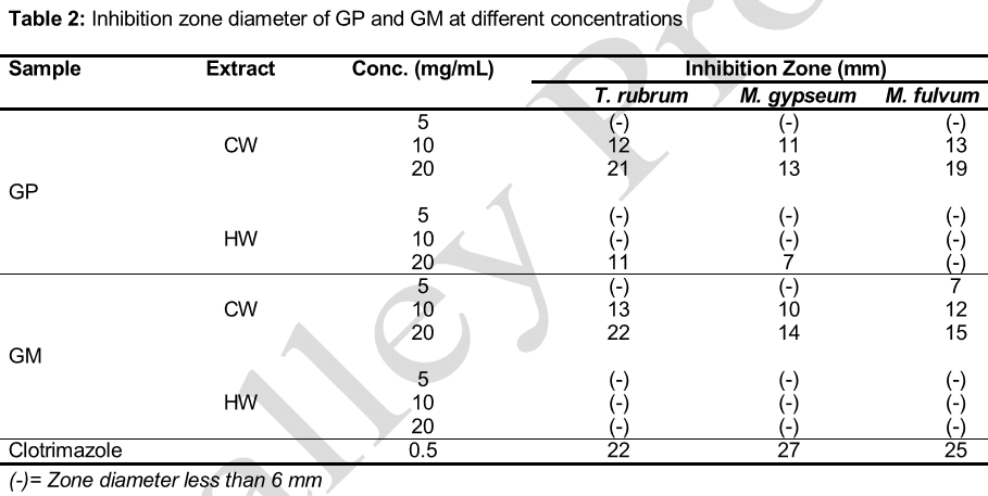
Agar-well diffusion method was employed for testing antifungal activity of the extracts of garcinia fruit. With a sterile cotton swab, 0.2 mL of fungal inoculum was spread evenly on the surface of the petri plate containing solidified SDA. Well of 6 mm diameter was made in the center of the agar plate with a sterile cork borer. The well was then filled with the respective extracts (0.3 mL) and allowed to diffuse at room temperature for 2 h. A control set was maintained with DMSO. Clotrimazole was used as a reference standard. The plates were then incubated at 28 ± 2 °C for 7-21 days depending on the growth rate of the test pathogens. The experiment was replicated thrice and the average results were recorded.
Mineral content analysis
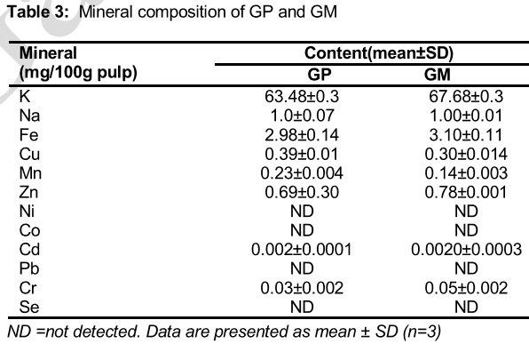
Mineral content was analyzed with Atomic Absorption Spectrophotometer, SIMADZU AAC-7000 analyzer. Dried pulp of GP and GM (100 mg) was digested with 3 mL concentrated nitric acid (65 %) and 0.25 mL hydrogen peroxide until a transparent solution was obtained. Finally after digestion the volume is made up to 30 mL with distilled water. The instrument was calibrated with known standards and samples analyzed at corresponding wavelengths. Na and K were determined by Flame photometer, ELICO CL378.
Statistical analysis
All assays were carried out in triplicate and the results expressed as mean ± SEM. Statistical analysis was carried out using Statistical Package for the Social Sciences (SPSS), version 16.0, and data analyzed using t-test, and p < 0.05 was considered statistically significant.
Results
TPC, TFC and antioxidant activities of GP and GM
TPC of CW and HW extracts of GP was 5.87 ± 0.06 and 4.45 ± 0.02 mg catechin equivalents/gm dry weight and in GM it was 5.46 ± 0.02 and 3.8 ± 0.3 mg catechin equivalents/gm dry weight respectively. It was thus revealed that the TPC in CW extracts was higher than that of HW extracts. TFC of CW and HW extracts of GP was found to be 5.61 ± 0.16 and 5.48 ± 0.05 mg quercetin equivalents/gm dry weight and in GM 3.69 ± 0.04 and 1.43 mg quercetin equivalents/gm dry weight respectively. TFC in both the extracts of GP were almost similar.
Similarly, in the case of DPPH, the IC50 values of the CW extracts were much lower (3.53 ± 0.04 and 1 ± 0.03 µg/mL) than than the standard ascorbic acid (5.56 ± 0.03 µg/mL) and it indicated that the GP and GM contained high antioxidant activity. Lower IC50 values indicated greater scavenging power. From the present results, it may be postulated that CW and HW extracts of GP and GM reduces the radical to corresponding hydrazine when it reacts with hydrogen donors in antioxidant principles. In case of H2O2, the IC50 values of CW extracts of GP and GM found to be 1.4 ± 0.02 and 1.44 ± 0.01 µg/mL respectively quite comparable with ascorbic acid (1 ± 0.03µg/mL) whereas higher values of IC50 values in case of HW extracts of both GP and GM indicates that some compounds destroyed during the boiling condition. Again CW and HW extracts of GM potentially inhibited the in vitro lipid peroxidation with an IC50 value of 30.36 ± 0.03 and 34.6 ± 0.03µg/mL against ascorbic acid 18 ± 0.03 μg/mL ().
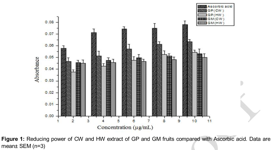
The reducing power of CW and HW extracts of GP and GM increased with increasing concentration. The antioxidant activities reported were concomitant with the development of reducing power. All the concentration of water extract showed significant activities when compared to standard ascorbic acid. The reducing power of the CW extract of GP and GM were higher than that of the HW extracts ().
Antifungal activity
At 20 mg/mL concentration, CW extract of GP and GM showed 19 mm and 15 mm zone diameter against Mycrosporum falvum (MF). At the same concentration, CW extract of GP and GM showed 13, 14 and 21, 22 mm zone diameter against Mycrosporum gypseum (MG) and Trichophyton rubrum (TR) respectively. But HW extract of GP and GM showed less activity. For this study a particular concentration (20 mg/mL) was considered, at which these fruits showed antifungal activity against some skin pathogenic fungi ().
Mineral content of GP and GM
showed that K, Na, Fe, Cu, Mn and Zn content of the samples were high in GP and GM but Ni, Co, Pb and Se were not detected. GP and GM were rich sources of Fe (2.98 ± 0.14 and 3.10 ± 0.11 mg/100g pulp).
Discussion
The emphasis on plant antioxidant has been increasing substantially in the recent times for the apprehension of some side effects of commercial antioxidants. The medicinal plants have different kinds of bioactive compounds, which have capabilities for termination of free radical chain reaction.
Phenolics and flavonoids are two main groups of polyphenols and both of these compounds are reported in fruits and vegetables. These two contents in fruits and vegetables can be influenced by species variety, environmental and growing conditions, maturity stages and harvesting factor [19-21]. Present study focused on the presence of phenolics and flavonoids in the dried pulp of GP and GM in CW and HW extracts of these two species separately. The study revealed that the TPC in the dried pulp of GP and GM fruit were found in higher amount in CW extracts than that of the HW extracts of both the fruits. Similarly, the CW extracts of GP and GM showed higher content of TFC. The present study clearly revealed that CW extracts are richer than HW extracts in TPC and TFC.
DPPH is a stable free radical, which was scavenged/neutralized by the extracts in a dose-dependent manner exhibiting high antioxidant potential of the extracts. Compounds with high antioxidant potential having high reduction capability of DPPH molecules. IC50 (amount of antioxidant present in the sample necessary to decrease the initial concentration by 50 %) value was calculated to determine the strength of antioxidant potential of the extracts. The lower the IC50 value, the higher is the antioxidant activity. CW extracts of GP and GM showed the lowest IC50 value in comparison to that of standard drug ascorbic acid in case of DPPH free radical scavenging activity.
H2O2 can cross cell membrane rapidly and produce hydroxyl radical, which can lead damage to the cell [22]. H2O2 probably reacts with Fe2+ and possibly Cu2+ ions to form hydroxyl radical, which may be the origin of many of its toxic effects [23]. So, it is necessary for cells to control the accumulation of H2O2 in the cells. In the present study, the CW extracts of both GP and GM scavenged the H2O2 radical very effectively and it was almost similar to that of the standard ascorbic acid. (). It is apparent from that the IC50 values of CW extracts of GP and GM against H2O2 free radical that CW extracts of both the species were much more effective which can be compared with the standard drug ascorbic acid.
The reducing capacity of a compound may serve as a significant indicator of its potential antioxidant activity [24]. The reducing power of the test sample always determined by the color changes of the test samples from yellow to green, due to the reduction of Fe3+/ferricyanide complex to the ferrous form and Fe2+ can be monitored by the measurement of the absorbance (OD value) at 700 nm [25]. CW extract showed very good reducing power than that of HW extracts.
Initiation of LPO by ferrous sulphate takes place through ferryl-perferryl complex [26]. The peroxidation of membrane lipids initiated by oxygen radical’s causes cardiovascular diseases and cancer [27]. This process may occur under enzymatic or non-enzymatic control. There has been an increasing interest in lipid peroxidation because formation of cytotoxic products such as MDA and 4-hydoxynonenal can influence cell function and the course of major human diseases [28]. In the present study, pink colour of MDA-TBA complex was detected at 532 nm and the inhibitory effect of CW and HW extracts of GP and GM on both ferric ion and ascorbic acid induced LPO on rat liver homogenate were calculated. The study showed that IC50 value of CW extracts of GP and GM against in vitro LPO is considerably low (), indicating that the extract are having excellent antioxidant potentiality.
Ethno medicines hold a great promise as a source of easily available effective antifungal agents particularly in developing countries, including India. Indigenous system of traditional medicine reports a number of plants for their antifungal efficacy [29]. The GP and GM extracts of the plant used in this study were found to be effective against the human dermatophytes. showed that CW extracts of GP and GM showed good antifungal activity.
Many antioxidant defenses depend on micronutrients. Some minerals are components of antioxidants enzymes: superoxide dismutase depends on Mn, Cu and Zn; catalase depends on Fe, and glutathione peroxidase on Se [30]. Of all minerals evaluated for GP and GM, K and Fe were detected in higher concentrations than other minerals. The ratio of K and Na plays an important role in maintaining the electrolyte balance of cells in the human body. Fe is an important part of hemoglobin for carrying oxygen to the body tissues. The availability of higher concentration of Fe in the samples indicates greater antioxidant properties.
Conclusion
The results obtained in the present study indicate that CW extracts of GP and GM exhibit higher reducing power, free radical scavenging and antifungal activity than HW extracts. There is thus some justification for the use by local tribes of NE region India, of the CW infusion of dried pulp of GP and GM against oxidative stress-related diseases. The findings of the present study suggest that GP and GM extracts are a potential source of natural antioxidant that may have great importance as therapeutic agents in preventing or slowing the progress of aging and oxidative stress related degenerative diseases.
Declarations
Acknowledgement
References
Archives


News Updates